Stroke

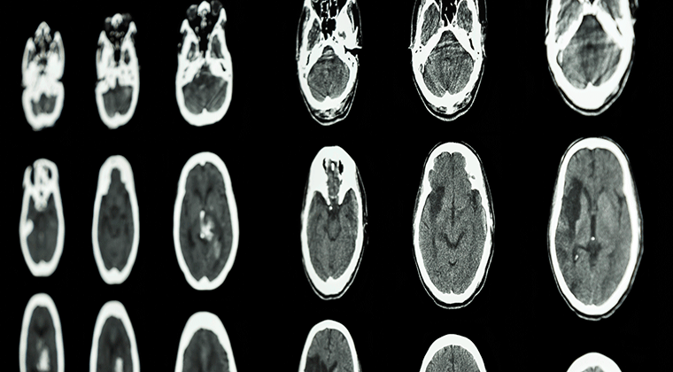
When something happens to the blood flow in the central nervous system, a cerebral vascular accident (CVA) or stroke occurs. A CVA most often occurs in the brain but, very rarely, it can also occur in the spinal cord. A stroke occurs when there is a change to blood flow in the central nervous system (CNS) due to damage to a blood vessel, rupture to the blood vessel or something stopping blood flow like a clot or emboli (globule of fat or other material or bubble of air.) Slowing of blood flow in the CNS can also lead to a stroke.
A person with loss of blood flow to the heart is said to be having a heart attack. The same process can occur in the central nervous system in either the spinal cord, brain or both. A person with loss of blood flow to the spine or brain or sudden bleeding in the spine or brain can be said to be having a “spine attack” or “brain attack.”
There are two main types of stroke:
- Ischemic strokes occur because of an obstruction (clot or emboli) within a blood vessel supplying blood to the brain or spinal cord. Obstructions typically result from plaque which has built up in the blood vessels that becomes dislodged and travels to the CNS. A clot or emboli typically originates in the heart, aorta or carotid arteries and travels to the CNS where it usually cannot pass through the tiny blood vessels. This stops blood flow to the affected part of the spinal cord or brain leading to stroke. A ischemic stroke from an unknown cause is called a cryptogenic stroke.
- A trans ischemic attack (TIA), sometimes called mini stroke, is an ischemic type of episode that resolves within 24 hours. A TIA is an indication that a stroke can follow. It can be a warning sign of stroke.
- Hemorrhagic strokes result from a blood vessel that ruptures and bleeds into the surrounding brain or spinal cord. The source of this type of stroke is hypertension, aneurism, arteriovenous malformation (AVM) or bleeding disorder.
- An arteriovenous malformation is a bleed that occurs where an artery meets a vein. The alignment of the artery transitioning to a vein is misaligned. These can occur anywhere in the body but are especially damaging when in the CNS (brain or spinal cord). AVMs are present at birth. Most go undetected without any issues unless they rupture.
When a CVA occurs, nerve cells in the affected area of the CNS can’t function due to a lack of oxygen. Surrounding tissue can be affected by an increase in swelling of the area. The spinal cord and brain are contained in rigid bones so swelling is constricted. Therefore, pressure within the CNS will compress soft nerve tissue.
Spinal cord CVA will result in complete (-plegia) or incomplete (-paresis) paralysis on both sides of the body depending on the level of injury in the spinal cord. Tetraplegia or tetraparesis (quadriplegia) involves the body and all four limbs. Paraplegia or paraparesis involves the lower part of the body. Effects of a spinal cord stroke can include alteration in mobility and function. Clot busting drugs do not seem to be effective in treating spinal cord stroke.
Brain CVA can affect the entire body, including paralysis or paresis (partial paralysis), cognitive and memory deficits, speech and visual issues, emotional difficulties, daily living challenges, and pain. Paralysis is a common outcome of stroke, often on one side of the body (hemiplegia). Paralysis may affect only the face, an arm or a leg, but most often, one entire side of the body and face is affected. A person who suffers a stroke in the left hemisphere (side) of the brain will show right-sided paralysis, or paresis. Likewise, a person with a stroke in the right hemisphere (side) will show deficits on the left side of the body.
Brainstem (the connection between the spinal cord and brain) CVA is a bit unique in that symptoms are vertigo and dizziness together with extreme imbalance. Double vision, slurred speech and decreased level of consciousness might be present. Brainstem stroke often mimics other neurological diseases. Diagnosis must be made early to take advantage of treatment.
Risk Factors and Symptoms
Stroke risk factors are divided by those you cannot control and those you can. If you have uncontrollable risk factors, do your best to control those you can to reduce risk.
Uncontrollable risk factors are those that are inherited and some pre-existing conditions. Included are age, gender, race, genetics and history of a prior TIA, stroke, or heart attack. People of all ages have strokes from newborns to the elderly, however as you get older, your risk of stroke increases due to changes in the body such as loss of artery elasticity (atherosclerosis). Women have a higher risk of stroke simply because they live longer than men. The stroke onset for women may be in these later years. Pregnancy complications, some birth control and post-menopausal hormones increase stroke risk. Genetics include family history of stroke or genetic diseases that can increase your risk of stroke. A person who has had a brain event such as TIA or stroke is at a much higher risk for a second one.
There are many factors that you can control to decrease your risk of stroke. You can greatly reduce your risk of stroke by controlling diseases that increase your risk. These diseases include high blood pressure, diabetes, obesity, high cholesterol, carotid artery disease, peripheral vascular disease, atrial fibrillation and other heart disease, and sickle cell disease. Many of these risk factors can be controlled by adopting a healthy diet and taking prescribed medication. Activity in your life will also help reduce your risk. Stopping smoking, alcohol and recreational drug use can significantly reduce the risk of a stroke.
If you have a spinal cord stroke or other spinal cord injury, the complication of Autonomic Dysreflexia (AD) puts you at higher risk for a stroke in your brain. AD is a medical emergency where blood pressure becomes elevated to excessive levels compared to the individual’s normal pressure. Over time, an individual with spinal cord injury will experience low blood pressure as their normal. An elevation to a normal range can indicate high blood pressure for that person. Find out more about Autonomic Dysreflexia (AD).
Some medical procedures have a risk for a spinal cord or brain stroke such as heart bypass surgery, open abdominal aortic aneurism, spinal surgery, or clot removal in the spinal arteries. Stroke risk is increased with any surgery, test or procedure that can create or dislodge a blood clot.
Stroke Symptoms
Spine Stroke
Stroke in the spinal cord has unclear symptoms. The signs of a spinal cord stroke can be confused with other illnesses. Any or all the symptoms can appear. The symptoms of a stroke in the spinal cord are:
- sudden and severe neck or back pain
- muscle weakness in the legs
- problems controlling the bowel and bladder (incontinence)
- feeling like there is a tight band around the torso
- muscle spasms
- numbness
- tingling sensations
- paralysis
- inability to feel heat or cold
Noticing a stroke in the spine can be difficult. You may have one, a combination or all the symptoms. It is important that you call 911 if your symptoms of a spinal cord stroke appear.
Brain Stroke
The symptoms of a stroke in the brain include:
F-FACIAL-drooping which might be noticeable on one entire side of the face or just in the eye or mouth
A-ARMS-an individual may not be able to raise one arm as high as the other or one arm may drift downward
S-SPEECH-may be slurry or strange as in use of odd words or sounds
T-TIME is of the essence as treatment needs to start immediately, call 911
You might have one of these symptoms, a few or all of them. You might not realize you are having the symptoms of a stroke. Someone else might notice these symptoms. Be sure to note the time of onset of symptoms. Write ‘onset: XX:XX am or pm’ on your body in case the stroke advances and you are unable to recall or speak when emergency personnel arrive. This is critical for treatment.
Results of Stroke
Results of a stroke depend on the location of the injury in the spinal cord or brain.
Spinal cord
Spinal cord stroke results in full or partial paralysis below the level of injury, bowel and bladder issues, sexual dysfunction, mobility and sensation difficulty. The result can be tetraplegia (quadriplegia), paraplegia, or one of the spinal cord syndromes. Abilities will vary depending on the location of the stroke in the spine.
After a stroke in the spinal cord, some may experience pain, uncomfortable numbness, or strange sensations. These may be due to many factors including damage to the spine which may interrupt the communication of messages between the body from the brain. Spinal cord stroke disrupts these messages at the level of the stroke in the spinal cord.
A stroke in the spinal cord can lead to depression due to the challenges of chronic illness. Loss of mental or physical function can be daunting. Changes in lifestyle and family dynamics compound the issue.
Brain
Left Brain Stroke can result in full or partial paralysis on the right side of the body. There might be difficulty in understand speech or speaking words, word finding or unusual use of words or sounds. The individual is typically cautious or even sometimes timid in behavior. Memory loss can be present.
Right Brain Stroke characteristics are full or partial paralysis on the left side of the body. There are often visual difficulties, quick and impulsive movements and thoughts. Memory loss can be present.
In left or right brain stroke, a visual field cut called hemianopsia can occur. The individual is not blind, but the visual field is reduced in both eyes. As a result, vision to one side is absent. The side of the visual field cut will be the same as side as paralysis in the body. The individual is unable to recognize that the visual field cut area is missing. This neglect can lead to injury to the neglected side even to the point that they do not recognize that body part as their own.
Brainstem
Brainstem stroke has the symptoms of vertigo and dizziness with imbalance. Functional abilities vary which can lead to locked in syndrome where the person understands what is happening but cannot physically or verbally respond. Memory loss can be present.
Your mental wellbeing can be affected by a stroke. Depending on the location of a stroke in the brain, you might have difficulty in remembering, being careless, irritable or have some confusion. Some people will laugh or cry inappropriately.
Treatment and Recovery
The appropriate response to a stroke is emergency action. Call 911. Do not attempt to drive yourself or let someone drive you to the hospital. Emergency personnel can begin the process of recovery. Every minute lost, from the onset of symptoms to the time of emergency contact, cuts into the limited window of opportunity for intervention.
Stroke prevention measures should be undertaken to reduce your risk of both a spine or brain attack. This includes the main three preventions; diet, exercise, smoking cessation. Other risk factors that you can control should be modified. Medication can also help reduce stroke risk. Medications to reduce cholesterol, blood thinners and antiplatelet medications should be taken as your healthcare provider prescribes.
Ischemic stroke is treated by removing the obstruction and restoring blood flow to the brain. This is done by breaking up the obstruction using the drug, tissue plasminogen activator (t-PA). Reversal of ischemic stroke can be achieved if the drug is administered within 6 hours of the onset of symptoms. The sooner the drug is administered, the better the outcome. Specially designated hospitals called, Stroke Centers, have streamlined assessments so t-PA can be administered in a timely fashion. You, or someone, must know the time of the onset of symptoms to be able to receive the drug.
Prior to hemorrhagic stroke, doctors attempt to prevent the rupture and bleeding of aneurysms and arteriovenous malformations. If the vessel has not ruptured, a ‘cage’ might be placed around the vessel to support the weakened area or the outpouching vessel might be ‘clipped’ and removed. If the vessel has already ruptured, surgery might be performed to remove the excess blood to the area, or the area might be monitored while the body reabsorbs the extra blood naturally.
Meanwhile, other neuroprotective drugs are being developed to prevent the wave of damage after the initial attack. Removing plaque and clots from the carotid arteries in a procedure called carotid endarterectomy, placing stents in the neck and brain blood vessels and blood thinning medications have all reduced the risk of stroke or especially, a second stroke.
General recovery guidelines show:
- 10 percent of stroke survivors recover almost completely
- 25 percent recover with minor impairments
- 40 percent experience moderate to severe impairments requiring special care
- 10 percent require care in a nursing home or other long-term care
- 15 percent die shortly after the stroke
Clinical practice guidelines are available which detail stroke treatment and rehabilitation strategies based on research evidence:
Adult Stroke Prevention:
Kernan WN, Ovbiagele B, Black HR, Bravata DM, Chimowitz MI, Ezekowitz MD, Fang MC, Fisher M, Furie KL, Heck DV, Johnston SC, Kasner SE, Kittner SJ, Mitchell PH, Rich MW, Richardson D, Schwamm LH, Wilson JA; American Heart Association Stroke Council, Council on Cardiovascular and Stroke Nursing, Council on Clinical Cardiology, and Council on Peripheral Vascular Disease. Guidelines for the prevention of stroke in patients with stroke and transient ischemic attack: a guideline for healthcare professionals from the American Heart Association/American Stroke Association. Stroke. 2014 Jul;45(7):2160-236. doi: 10.1161/STR.0000000000000024. Epub 2014 May 1. Erratum in: Stroke. 2015 Feb;46(2):e54.
Adult Stroke Rehabilitation:
Duncan PW, Zorowitz R, Bates B, Choi JY, Glasberg JJ, Graham GD, Katz RC, Lamberty K, Reker D. Management of Adult Stroke Rehabilitation Care: a clinical practice guideline. Stroke. 2005 Sep;36(9): e100-43.
Pediatric, Neonatal:
Debillon T, Ego A, Chabrier S. Clinical practice guidelines for neonatal arterial ischaemic stroke. Dev Med Child Neurol. 2017 Sep;59(9):980-981. doi: 10.1111/dmcn.13451.
Rehabilitation
Rehabilitation starts immediately after a stroke of the spine or brain and should be continued as necessary after release from the hospital. Rehabilitation options may include the rehab unit of a hospital, a subacute care unit, a rehab hospital, home therapy, outpatient care, or long-term care in a nursing facility. Your ability to participate in therapy will determine the level of care appropriate for your rehabilitation. Your healthcare professional, case manager or social worker in the acute care facility will help you determine the level of care that is appropriate.
The goal in rehabilitation is to improve function so that an individual can become as independent as possible. This must be accomplished in a way that preserves dignity while motivating the individual to relearn basic skills such as eating, dressing and walking. Rehabilitation builds strength, capability, and confidence so a person can continue daily activities despite the effects of stroke.
Activities may include the following:
- Self-care skills such as feeding, grooming, bathing, and dressing
- Mobility skills such as transferring, walking, or moving a wheelchair
- Communication skills; cognitive skills such as memory or problem-solving
- Social skills for interacting with other people.
- Constraint Induced Therapy
- Functional Electrical Stimulation
- Partial Weight Supported Walking
- Activity Based Exercise
- Aquatic Therapy
- Music Therapy
After stroke, individuals often find that once-simple tasks around the house become extremely difficult or impossible. Many adaptive devices and techniques are available to help people regain their independence and function safely and easily. The home usually can be modified so the individual can manage personal needs.
An approach called constraint-induced movement-based therapy (CIT) has improved recovery in people who have lost some function in a single limb. The therapy entails immobilizing a patient’s unaffected limb to force use of the affected limb. CIT is thought to promote a remodeling of nerve pathways, or plasticity.
Technological advancement has provided therapies that are being incorporated into rehabilitation care. Electronic devices that allow speech by using a pointer, eye movement or even brain waves are available in many varieties which can be selected for specific individual needs.
Newer, advanced therapies have been added to the physical rehabilitation program to treat stroke. Electrodes placed on the skin with use of electrical stimulation can provide input to nerves affected in the body. Machines that provide repetitive movement such as partial weight supported walking, ‘gloves’ that create hand movement and activity-based therapy provide stimulation to limbs for return of function. Mirror therapy allows the individual to look at the affected side of their body to learn to gain control with the same side of the brain. Aquatic therapy allows the body to move using the buoyancy of the water. Music therapy connects rhythm to movement. All can enhance recovery from stroke.
Recovery from a stroke can take time. At first, recovery may seem to be on a fast trajectory as swelling and trauma to the brain resolve. Over time, return of function may seem to slow but recovery is still occurring. It is a myth that the central nervous system does not attempt to recover. It is a myth that the nerve cells you are born with are the only ones you will have.
The nervous system is capable of great improvement. It wants to repair itself and restore function through a process called plasticity. New nerve cells are being developed throughout your life. Keeping up with your home exercises will help continue improvement, although at a slower rate.
Facts and Figures
Stroke is the nation’s fourth leading cause of death and is a leading cause of serious, long-term disability in the United States. Just a few years ago, it was the third highest cause of death. The survival rate is increasing due to the advancements in treatments.
Spinal stroke is estimated to occur in 54 out of 1 million people. Spinal stroke is rare. Therefore, general stroke information is provided.
Approximately, 6 million individuals who have had a stroke are alive today.
800,000 strokes occur in the US every year.
87% of strokes in the brain are ischemic. 13% of strokes in the brain are hemorrhagic.
Individuals with atrial fibrillation (abnormal heartbeat) have a 5 times greater chance of stroke.
World Stroke Day is October 29th.
Eighty percent of strokes are preventable by controlling your blood pressure, cholesterol and blood sugar. Also, eating a healthy diet, losing weight and being active. Stop smoking. Take low dose aspirin only if recommended by your healthcare professional. (Taking aspirin when not needed can damage your health in other ways.)
Source: National Institutes of Health (NIH)
Research
Research advances have led to new therapies and new hope for people who are at risk or who have had a stroke. There are literally thousands of research studies being conducted on various aspects of stroke. The study of stroke prevention, control of risk factors and treatments are underway.
The uncontrollable factors that increase risk of stroke are being studied in the laboratory. Genetic therapy is being studied to modify these factors. One interesting approach is to slow blood flow to the brain as animals who hibernate do. Another is using hypothermia or extreme cooling of the body to reduce the need for blood flow. On the other end of study is to increase blood flow to the brain through transcranial stimulation, an external device to increase blood flow to the affected part of the brain or spinal cord after a stroke.
When a stroke occurs, some spinal cells die immediately; others remain at risk for hours and even days due to an ongoing sequence of destruction. Some damaged cells can be saved by early intervention with neuroprotective drugs. These drugs that work in a variety of ways are in clinical trials.
Clinical trials are underway. These are research studies performed with people. Use of stents in the carotid arteries (much like in the heart) are shown to be as effective as surgery. Medical treatment of taking aspirin with and without antiplatelet medication is being studied as a treatment to reduce risk for a second stroke. Other medications to control albumin and glucose are being studied.
Stem cells are being studied as a possible treatment for just about every diagnosis. These studies are in the early phases. There is not a cure for stroke using stem cells at the present time.
Resources and Support for Managing Strokes
If you are looking for more information on strokes or have a specific question, our Information Specialists are available business weekdays, Monday through Friday, toll-free at 800-539-7309 from 9:00 am to 8:00 pm ET.
Additionally, the Reeve Foundation maintains a fact sheet on how to support individuals living with a stroke with additional resources from trusted Reeve Foundation sources. Check out our repository of fact sheets on hundreds of topics ranging from state resources to secondary complications of paralysis.
We encourage you to reach out to stroke support groups and organizations, including:
- American Stroke Association (ASA)
- Stroke Family Support Network
- National Stroke Association (NSA)
- National Heart Association
- National Institute of Neurological Disorders and Stroke
Be sure to check with the above national organizations for chapters and resources in your local community.
Further Reading
Stroke Symptoms—Spine section:
Hill VA, Towfighi A. Modifiable Risk Factors for Stroke and Strategies for Stroke Prevention. Semin Neurol. 2017 Jun;37(3):237-258. doi: 10.1055/s-0037-1603685. Epub 2017 Jul 31.
Alloubani A, Saleh A, Abdelhafiz I. Hypertension and diabetes mellitus as a predictive risk factors for stroke. Diabetes Metab Syndr. 2018 Jul;12(4):577-584. doi: 0.1016/j.dsx.2018.03.009. Epub 2018 Mar 19.
Results of Stroke section:
Hankey GJ. Stroke. Lancet. 2017 Feb 11;389(10069):641-654. doi: 10.1016/S0140-6736(16)30962-X. Epub 2016 Sep 13.
Frolov A, Feuerstein J, Subramanian PS. Homonymous Hemianopia and Vision Restoration Therapy. Homonymous Hemianopia and Vision Restoration Therapy. Neurol Clin. 2017 Feb;35(1):29-43. doi: 10.1016/j.ncl.2016.08.010.
Bartolomeo P, Thiebaut de Schotten M. Let thy left brain know what thy right brain doeth: Inter-hemispheric compensation of functional deficits after brain damage. Neuropsychologia. 2016 Dec;93(Pt B):407-412. doi: 10.1016/j.neuropsychologia.2016.06.016. Epub 2016 Jun 14.
Treatment and Recovery section:
Siegel J, Pizzi MA, Brent Peel J, Alejos D, Mbabuike N, Brown BL, Hodge D, David Freeman W. Update on Neurocritical Care of Stroke. Curr Cardiol Rep. 2017 Aug;19(8):67. doi: 10.1007/s11886-017-0881-7
Hadidi NN, Huna Wagner RL, Lindquist R. Nonpharmacological Treatments for Post-Stroke Depression: An Integrative Review of the Literature. Res Gerontol Nurs. 2017 Jul 1;10(4):182-195. doi: 10.3928/19404921-20170524-02. Epub 2017 May 30
Rehabilitation section:
Ware R, Moore M. Validity of measures of neurological status used for predicting functional independence in adults after a cerebrovascular accident: a systematic review protocol. JBI Database System Rev Implement Rep. 2017 Mar;15(3):603-606. doi: 10.11124/JBISRIR-2016-002978.
Pin-Barre C, Laurin J. Physical Exercise as a Diagnostic, Rehabilitation, and Preventive Tool: Influence on Neuroplasticity and Motor Recovery after Stroke. Neural Plast. 2015;2015:608581. doi: 10.1155/2015/608581. Epub 2015 Nov 23. Review.
Hara Y. Brain plasticity and rehabilitation in stroke patients. J Nippon Med Sch. 2015;82(1):4-13. doi: 10.1272/jnms.82.4. Review.
Facts and Figures section:
Thrift AG, Thayabaranathan T, Howard G, Howard VJ, Rothwell PM, Feigin VL, Norrving B, Donnan GA, Cadilhac DA. Global stroke statistics. Int J Stroke. 2017 Jan;12(1):13-32. doi: 10.1177/1747493016676285.
Research section:
Hu X, Tong KY, Li R, Chen M, Xue JJ, Ho SK, Chen PN. Combined functional electrical stimulation (FES) and robotic system for wrist rehabiliation after stroke. Stud Health Technol Inform. 2010;154:223-8.
Russo MJ, Prodan V, Meda NN, Carcavallo L, Muracioli A, Sabe L, Bonamico L, Allegri RF, Olmos L. High-technology augmentative communication for adults with post-stroke aphasia: a systematic review. Expert Rev Med Devices. 2017 May;14(5):355-370. doi: 10.1080/17434440.2017.1324291. Epub 2017 May 9.
Hoermann S, Ferreira Dos Santos L, Morkisch N, Jettkowski K, Sillis M, Devan H, Kanagasabai PS, Schmidt H, Krüger J, Dohle C, Regenbrecht H, Hale L, Cutfield NJ. Computerised mirror therapy with Augmented Reflection Technology for early stroke rehabilitation: clinical feasibility and integration as an adjunct therapy. Disabil Rehabil. 2017 Jul;39(15):1503-1514. doi: 10.1080/09638288.2017.1291765. Epub 2017 Mar 3.
Bertani R, Melegari C, De Cola MC, Bramanti A, Bramanti P, Calabrò RS. Effects of robot-assisted upper limb rehabilitation in stroke patients: a systematic review with meta-analysis. Neurol Sci. 2017 Sep;38(9):1561-1569. doi: 10.1007/s10072-017-2995-5. Epub 2017 May 24.

